Figure 6 from Femoral Hernia: A Review of the Clinical Anatomy and
By A Mystery Man Writer
Description
Figure 6. Femoral hernia repair in clean operation. (a) The narrow side of the mesh is sutured to Cooper’s ligament; (b) The mesh is sutured to the iliopubic tract or shelving portion of the inguinal ligament; (c) The posterior wall of the inguinal canal is reinforced, as in Lichtenstein’s repair. - "Femoral Hernia: A Review of the Clinical Anatomy and Surgical Treatment"
Myopectineal orifice. The oval-shaped myopectineal orifice (green

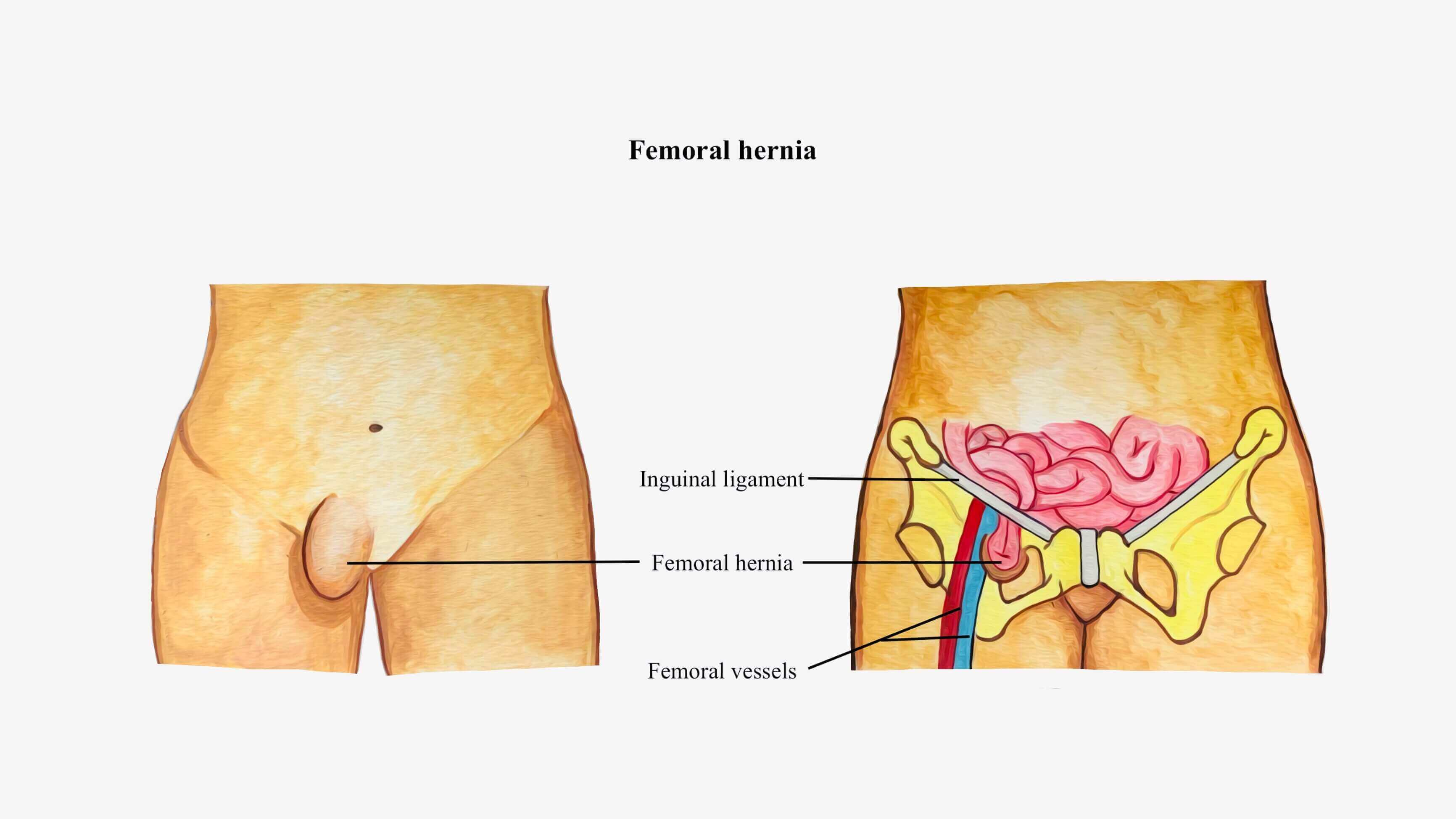
Femoral hernia: Symptoms, pictures, treatments, and more

Figure 12 from Femoral hernia repair.
Femoral Hernia - A Review of Clinical Anatomy

Femoral Hernia - A Review of Clinical Anatomy

Femoral Hernia: A Review of the Clinical Anatomy and Surgical Treatment

Femoral Hernia - A Review of Clinical Anatomy

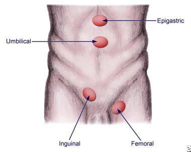
Figure, Abdominal Hernias Contributed by T Silappathikaram] - StatPearls - NCBI Bookshelf
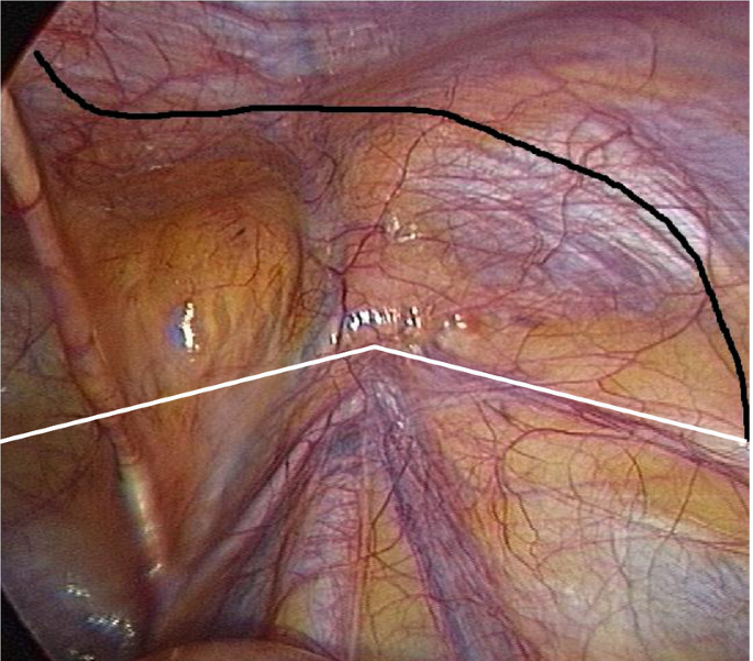
Clinical Anatomy of the Groin: Posterior Laparoscopic Approach
Hernia Reduction: Background, Indications, Contraindications

From inguinal to giant femoral hernia: An unusual postoperative twist - A rare case report - ScienceDirect

Embryonic developmental process and clinical anatomy of the preperitoneal fascia and its clinical significance

Clinical Anatomy of the Groin: Posterior Laparoscopic Approach

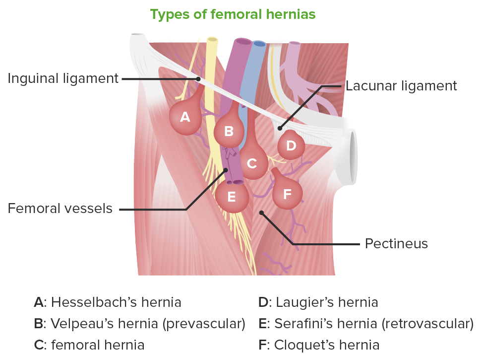
Schematic view of right femoral region illustrating variants of femoral
from
per adult (price varies by group size)