LMs of living cells showing chloroplasts. Fig. 1. Girdle view of
By A Mystery Man Writer
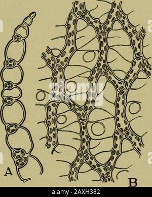
Description

LMs of auxospores and initial cells. Arrowheads indicate mother

PDF) Fine-structure of the vegetative frustule, perizonium and
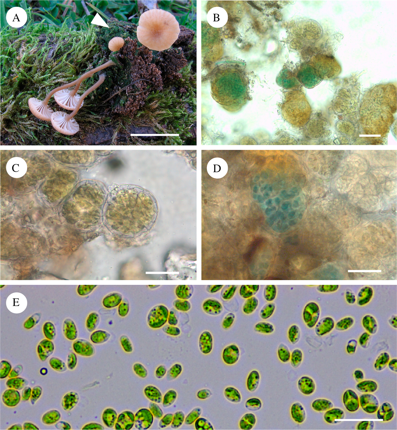
Symbiont composition of the basidiolichen Lichenomphalia

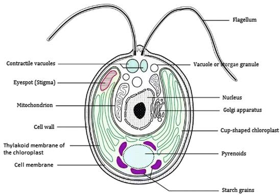
Diagrams to show arrangement of chloroplasts within cell, shown in
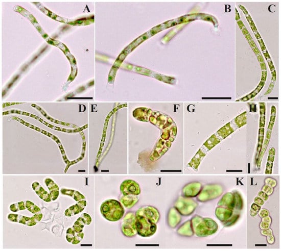
Nature and development of plants . Fig. 6. Surface view of

Masahiko Idei's research works Bunkyo University, Tokyo and

Chloroplasts in a living mesophyll cell of spinach observed by

The Walter Reed Visual Assessment Scale (used with permission from

Medians of WRVAS questions for each curve pattern

The Walter Reed Visual Assessment Scale (used with permission from

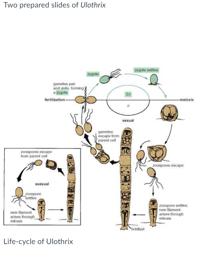
Classification of Plant-like Protists - Advanced ( Read
from
per adult (price varies by group size)