Optical Coherence Tomography: Imaging Mouse Retinal Ganglion Cells In Vivo
By A Mystery Man Writer
Description
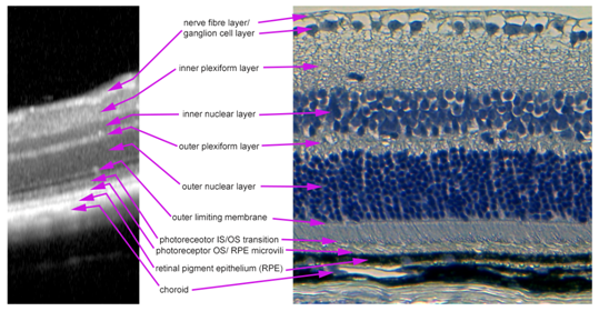
Scientific Article | Structural changes in the retina are common manifestations of ophthalmic diseases.
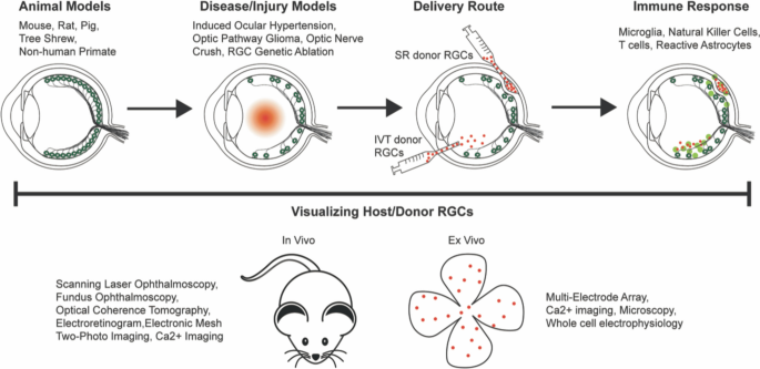
Retinal ganglion cell repopulation for vision restoration in optic neuropathy: a roadmap from the RReSTORe Consortium, Molecular Neurodegeneration
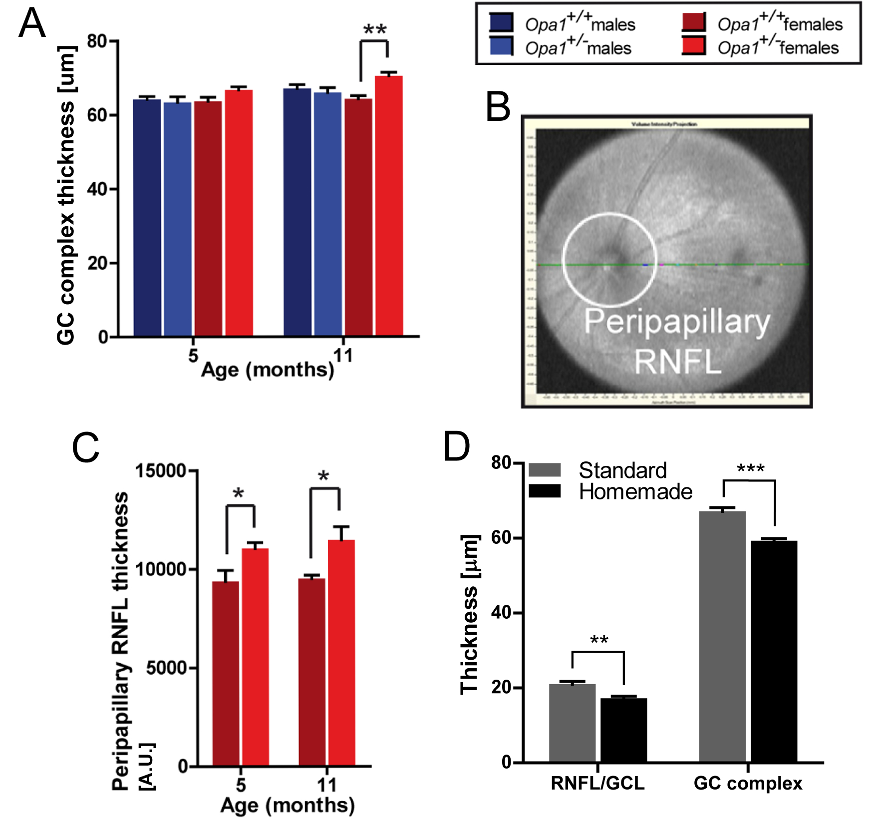
Longitudinal In Vivo Imaging of Retinal Ganglion Cells and Retinal Thickness Changes Following Optic Nerve Injury in Mice

Genes, Free Full-Text

All Protocols and Video Articles in JoVE

Adaptive optics with combined optical coherence tomography and

Monitoring retinal changes with optical coherence tomography predicts neuronal loss in experimental autoimmune encephalomyelitis, Journal of Neuroinflammation

Human adipose tissue-derived stem cell extracellular vesicles attenuate ocular hypertension-induced retinal ganglion cell damage by inhibiting microglia- TLR4/MAPK/NF-κB proinflammatory cascade signaling, Acta Neuropathologica Communications
Quantification of optical In-Vivo imaging of retinal and choroidal neovascularisation and cell migration in the mouse fundus - MedCrave online

Transplanted human induced pluripotent stem cells- derived retinal ganglion cells embed within mouse retinas and are electrophysiologically functional - ScienceDirect

Optical Coherence Tomography: Imaging Mouse Retinal Ganglion Cells In Vivo
Optical Coherence Tomography: Imaging Mouse Retinal Ganglion Cells In Vivo

PDF] In vivo imaging and counting of rat retinal ganglion cells using a scanning laser ophthalmoscope.

Topical Nerve Growth Factor (NGF) restores electrophysiological alterations in the Ins2Akita mouse model of diabetic retinopathy - ScienceDirect
from
per adult (price varies by group size)