Medial view of left knee region highlighting various fascial
By A Mystery Man Writer
Description
Download scientific diagram | Medial view of left knee region highlighting various fascial components surrounding the semitendinosus muscle. From the superficial to the deep aspect: the fascia lata, the paratenon and the epimysium from publication: Anatomical study of paratenons and fascia lata connections in the posteromedial knee region | Introduction In the last decade, fascia research increased significantly in various aspects such as anatomical and biomechanical features related to epimuscular force transmission. Methods The present anatomic study focuses on macroscopic observations of the potential | Fascia Lata, Hamstring muscles and Fascia | ResearchGate, the professional network for scientists.
Middle ear: Anatomy, relating structures and supply

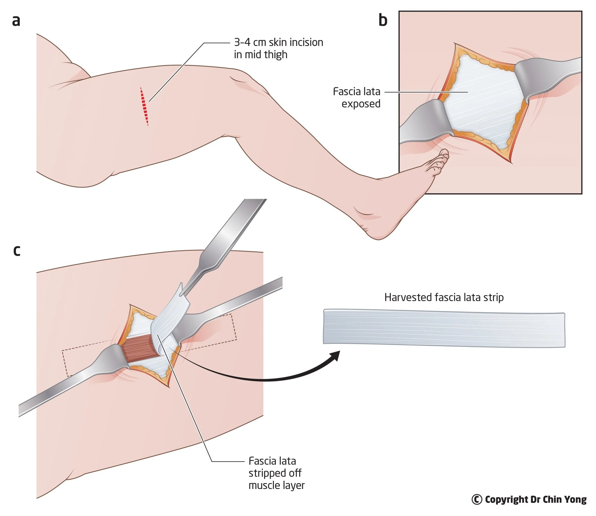
Key Surgically Relevant Anatomy of the Medial and Lateral Aspects

AP and lateral radiographs of the left knee demonstrate lucency
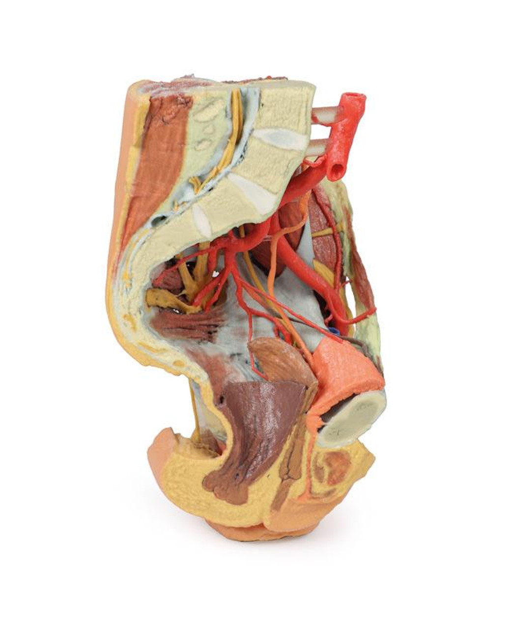
3D Printed Female Left Pelvis and Proximal Thigh

Anatomical study of paratenons and fascia lata connections in the posteromedial knee region

Muscles of the Posterior Leg - Attachments - Actions - TeachMeAnatomy

Benoit BEYER, Assoc. Prof., PT, MSc, PhD, Université Libre de Bruxelles, Brussels, ULB, Faculty of Motricity Sciences (FMS)
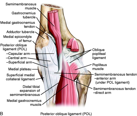
Medial and Anterior Knee Anatomy
Superior tibial plateau view of the Medial and Lateral Meniscus as well as the anterior and posterior cruciate ligament. Bent knee view of the right

Normal Left Knee Anatomy - Superior tibial plateau, sagittal & anterior view
from
per adult (price varies by group size)