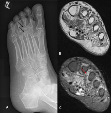
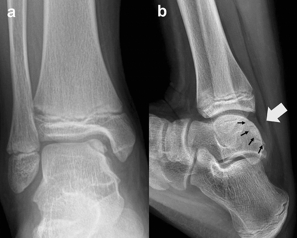
Foot X-ray of a 10 year-old male patient (white arrow indicates
By A Mystery Man Writer
Description
Gluteus Minimus Anatomy and Tear Patterns
The Homeostasis of Cartilage Matrix Remodeling and the Regulation of Volume-Sensitive Ion Channel

Cervical adjacent segment disease: Risks and complications following cervical fusion – Caring Medical Florida

Example of a detached house.
Complex Regional Pain Syndrome Type 1 (Reflex Sympathetic Dystrophy) Imaging: Practice Essentials, Radiography, Positron Emission Tomography (PET)

Example of a detached house.

Plain film radiograph of 20-year-old patient with foot pain associated

X ray of the left foot of a 29 years old male patient. Normal, Stock Photo, Picture And Rights Managed Image. Pic. BSI-BSIP-013095-065
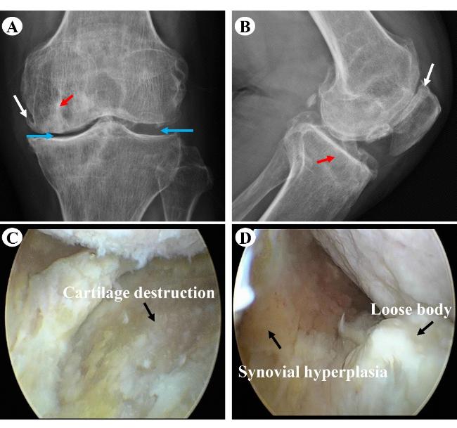
Second-look arthroscopic and magnetic resonance analysis after internal fixation of osteochondral lesions of the talus

X-ray images of an ankle with bony anterior ankle impingement
from
per adult (price varies by group size)